

Retinal & Interior Imaging
Advanced eye imaging is the key to assessing and maintaining optimal eye health
We are so lucky at Blink to be able to offer advanced eye imaging through the use of our Optomap, Optical Coherence Tonographer and slit lamp cameras.
With the development of technology, eye examinations have advanced over the years. One of the most significant advancements is the introduction of advanced retinal imaging and anterior eye imaging. Being able to accurately check your eye health as well as test your vision is essential for a comprehensive eye examination. This is why we’ve invested in Optomap Ultra-Widefield Imaging, the Optical Coherence Tonography Scan and the DC4 Slit lamp camera. These devices allow us to be more thorough in examining your eyes.
Why is retinal imaging so important?
Retinal imaging is an important diagnostic tool used to evaluate the health of the retina, which is the part of the eye responsible for transmitting visual information to the brain. Here are some reasons why retinal imaging is so important:
Retinal imaging allows us to identify eye diseases such as macular degeneration, glaucoma, and diabetic retinopathy in their early stages when they are more treatable.
Retinal imaging is a key component of a comprehensive eye exam, which can detect and diagnose eye conditions that may not have any noticeable symptoms.
Retinal imaging provides a baseline of the eye's condition, which can be used to track changes over time and monitor the effectiveness of treatment.
Retinal images can help us explain eye conditions to our patients and provide visual evidence of any changes or progress over time.
Retinal imaging is a non-invasive and painless procedure that can be performed quickly and easily and give us instant results
Overall, retinal imaging is an important tool in the diagnosis, monitoring, and treatment of eye diseases, and can help improve the overall health and vision of patients. We are delighted to be able to offer these services at Blink.

How does retinal imaging work?
Optomap
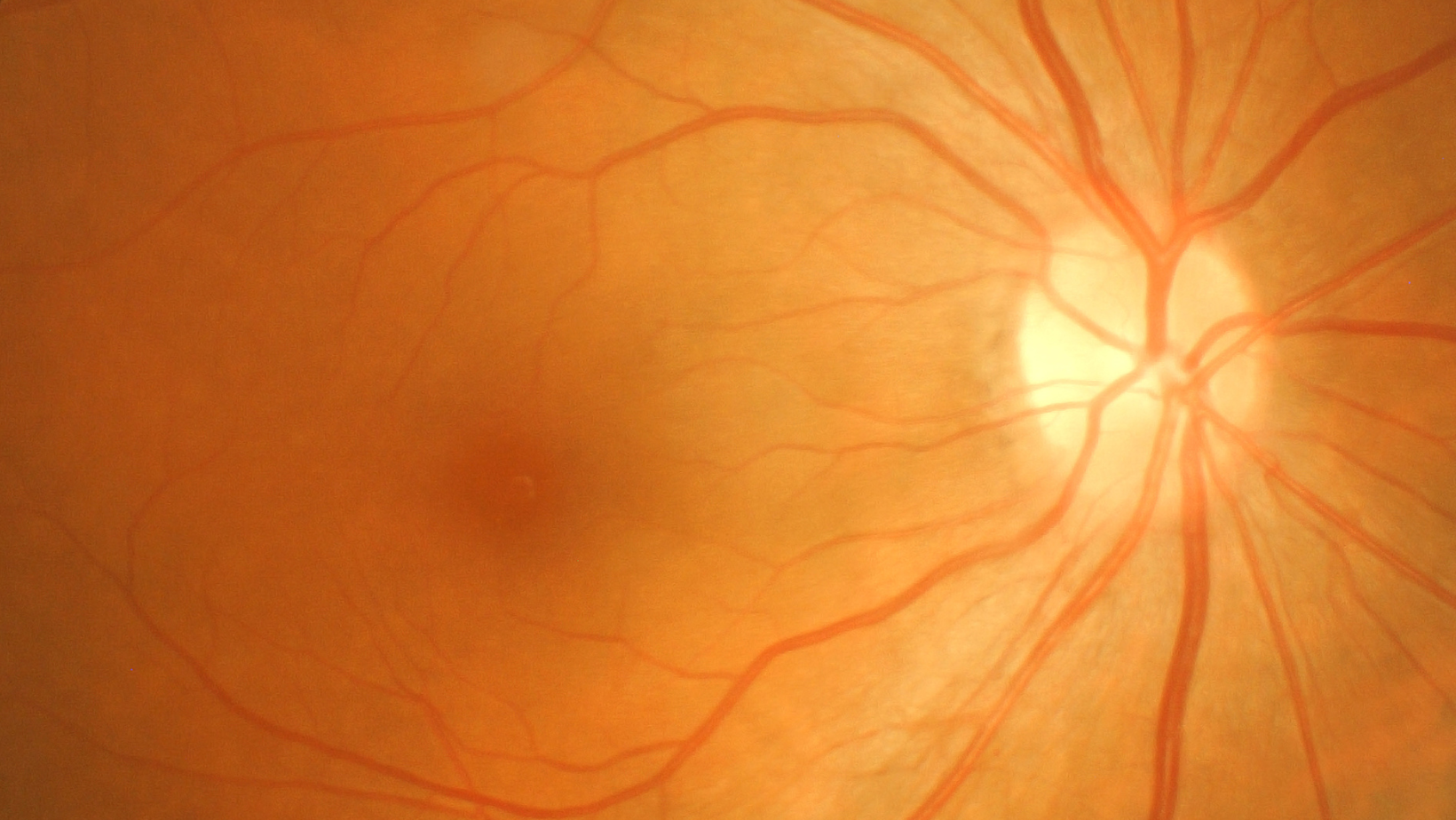
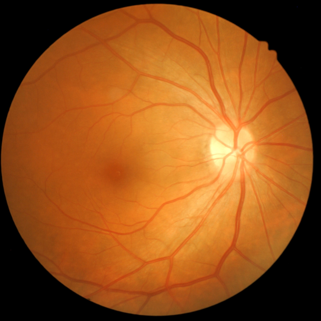
The Optomap ultra-widefield scan captures and produces a 200° panoramic digital image that covers up to 87% of your retina in less than half a second. Traditional imaging only allowed us to see 15% of your retina. The Optomap scan allows us to look at different layers to have a more in depth view.

Optical Coherence Tonography (OCT)
Our Optomap ultra-widefield technology is complemented by the Topcon OCT scan. This OCT scan is the equivalent of a CAT scan for your eyes, allowing us to produce 3D images that show a cross-section of the layers which make up your retina, allowing our optometrists to, quite literally, see beneath the surface. The software for both scans enable us to monitor changes over the years when we do comparative scans.
Both scans are non-invasive, quick, easy and painless. Within seconds, all images will be taken, producing instant results that we can analyse and discuss with you.
These advanced scans are not covered by the NHS. We will be able to discuss and demonstrate these scans with you at your eye examination.

Anterior Eye Imaging - Slit lamp camera
A slit lamp camera is a specialised optical imaging device used by us to capture high-quality images of the anterior eye.
The slit lamp camera is commonly used in practices for diagnostic purposes. The images captured by the camera can also be used to educate patients on their eye health and provide visual documentation of changes over time.
Our Slit lamp camera also gives us access to teleophthalmology. This provides remote ophthalmic care to patients. It involves the use of digital technology to remotely diagnose and treat eye-related conditions. This allows the Optometrists at Blink to access Ophthalmologists advice - allowing us to share (with your consent) images/scans and we can even have a three way consultation with an Ophthalmologist if thought beneficial.